CASES BY HENK
Non-Surgical Endodontics
Endodontic Microsurgery
The broad range of challenging cases shown here are a few of the many thousands that Henk has performed.
They demonstrate the complexity of root canal anatomy as well the potential for bony healing to occur, even with very large lesions. Henk embraced the need for CBCT in endo long ago, and many cases demonstrate his use of 3D imaging.
NON-SURGICAL CASES
Severely Curved Roots
CASE 1
The challenges of treating this upper 6 with curved roots and calcified pulp and canals were identified by the referring dentist. Henk performed treatment and placed an amalgam core. The six month review radiograph with the tooth crowned, is shown.
CASE 2
RCT was attempted on this upper 6. The extreme curvatures could not be negotiated and the patient was referred to Henk who performed treatment and restored the access cavity with a reinforced bonded resin. The patient was returned to the dentist for a definative cuspal protection restoration.
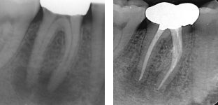
Deep Canal Divisions
CASE 1
This maxilliary first molar had a wide, blunt palatal root and this can hint at the presence of complex anatomy. A CBCT was taken and the sagittal slice clearly shows the anatomy that was duplicated on the final radiograph. The distal canal had restrictive access apically, only allowing for instrumentation with small hand files and hydraulic obturation with sealed (SystemB).
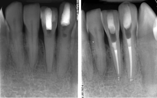
CASE 2
The lower premolars can be very challenging to treat as they comonly have deep canal divisions. This LR5 was referred as RCT had been attempted. Henk shaped, cleaaned and obturated three canals in the apical third. A bonded resin core was placed for the referring dentist to prepare for a cuspal protection restoration
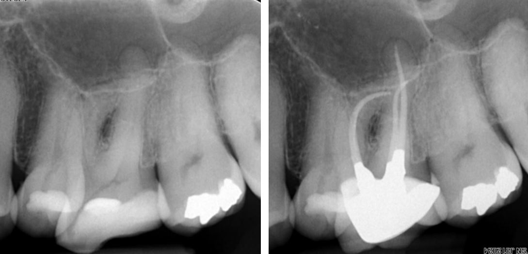
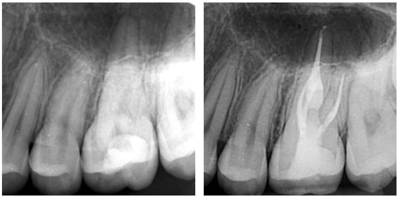
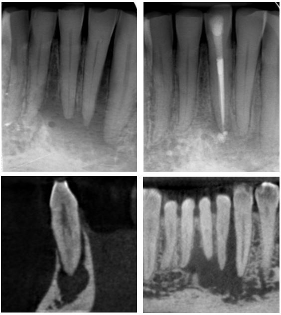
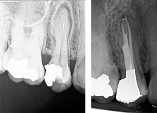
CASE 3
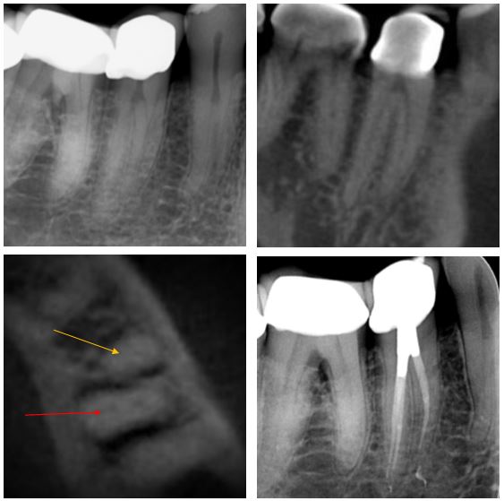
This UR6 developed symptomatic pulpitis secondary to a cracked cusp.The clinical picture was complex and a CBCT scan was used to confirm diagnosis. This showed deep bifurcation of the palatal canal. The axial slice section has arrows highting the mesial and distal branches of the root. Bioceramics were used to obturate, therfore not terribly radiopaque. A base of GIC was placed under a bonded resin core, ready for referring dentist to prep.
Unusual Root Canal Configurations
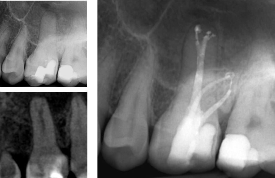
CASE 1
Lower premolars can have very challenging anatomy. This recently crowned LR5 developed an irriversable pulpitis and the pre-treatment radiograph hints at it having a mesial and distal root. A CBCT was taken and the sagittal slice (top right) confirms two seperate roots. The axial slice with yellow arrow at mesial root shows two canals in the distal root (red arrow). Following treatment through the crown an amalgam access cavity restoration was placed for shear resistance.
CASE 2
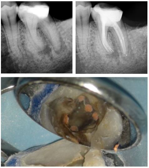
A heavily restored LL6 was referred as it required a crown and had a draining fistula associated with the large periapical radiolucencies. Five canals (three mesial and two distal) were cleaned and dressed with calcium hydroxide for six weeks. On completion of the case the pulp chamber was cleaned with microabrasion and a bonded core was placed for the referring dentist to prepare. The final radiograph shows periapical healing underway. The filled canal orrifices are shown on the clinical radiograph.
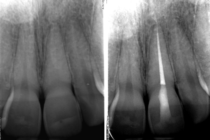
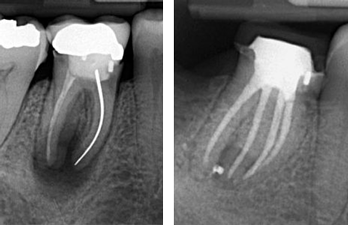
Teeth With Very Long Roots
CASE 1
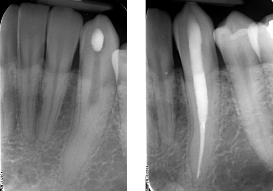
This 32mm long lower canine shows unusual and sudden canal obliteration at mid-root level. Deep ultrasonic excavation to 20mm allowed for canal catheterization and completion of treatment.
The access cavity was restored with Fuji IX GIC.
CASE 2
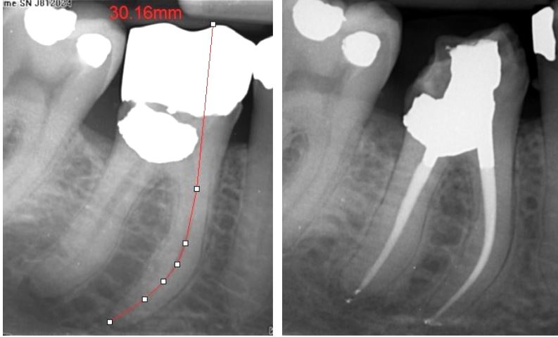
A crowned lower 6 with pre-treatment estimate length of 30mm. Secondary caries indicated crown removal and replacement following RCT. The large class V buccal amalgam had been placed in the bucccal canal orrifices making this case more challenging. A temporary crown was placed and a the six mont review radiograph shows periapical healing so that a new crown can be placed
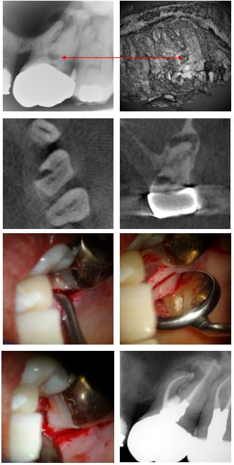
Cervical Root Resorption Treated With Aid Of CBCT
Case 1 – Molar Tooth
This gentleman presented with discomfort from the left hand side posterior teeth. A periapical radiograph showed periapical radiolucencies associated with UL6 and UL7. A cervical radiolucency, typical of cervical root resorption was also obvious. This indicated CBCT investigation and the associated 3D rendered image and corresponding lesion indicated by the red arrows. The next two CBCT slices are the axial and coronal viers of the lesion.
The tooth was root canal system was shaped, cleaned and dressed with calcium hydroxide and the access cavity filled with a temporary material. The clinical photos show the surgically exposed lesion prior to, and following curretage. The defect was restored with Geristore®, a resin-ionomer. On a second visit the root canal was completed and the access cavity restored with amalgam for shear resistance.
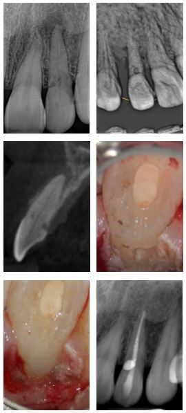
CASE 2
This unrestored UL2 developed a periapical abscess. The referring dentist noticed a small cervical radiolucency and referred to Henk for management. Cervical root resorption was suspected and confirmed with CBCT. Following RCT, a palatal flap was raised and the defect cleaned and restored with Geristore®. A six month review radiograph confirms periapical healing (bottom right)
External Apical Resorption
CASE 1
This LR7 has a severely resorbed distal root likely mediated by pressure from the impacted 8 (surface resorption) combined with inflamatory resorption typical of teeth with chronic periapical disease. An OPG was taken to rule out presence of a dentigerous cyst and this showed the same clinical picture at LL7 (top right).
Definative treatment required RCT and removal of the 8. Bottom right shows Post treatment RCT and core placement. Bottom right is six month follow-up radiograph once referring dentist had placed a crown, it shows good periapical healing preceding extraction of LR8.
CASE 2
A more typical case of inflamatory external root resorption due to chronic periapical inflamation. As the periapical inflamation is an extension of pulpal disease, conventional endodontic management leads to good outcomes.
This crowned LR6 had secondary caries requiring crown and caries removal. Following RCT, a core and tempory crown was placed. A three month review radiograph shows good healing underway so that referring dentist can place a new crown.

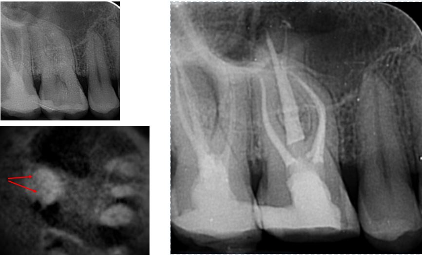
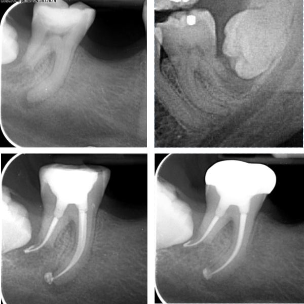
Internal Resorption
CASE 1
This root treated upper 6 had an associated buccal draining fistula and large periapical radiolucency on the mesial root. The mesial root has a mid-root radiolucent that indicated a CBCT scan for resorption diagnosis. The sagittal and axial slice are shown. The axial slice with red arrow at the mesial root shows frank root perforation has not occured. A modified cleaning protocol incorporating ultrasonic file activation was used in the mesial canal system and the case was obturated with bioceramics prior to placing a GIC base and bonded composite core. The six month review radiograph shows excellent periapical healing underway.
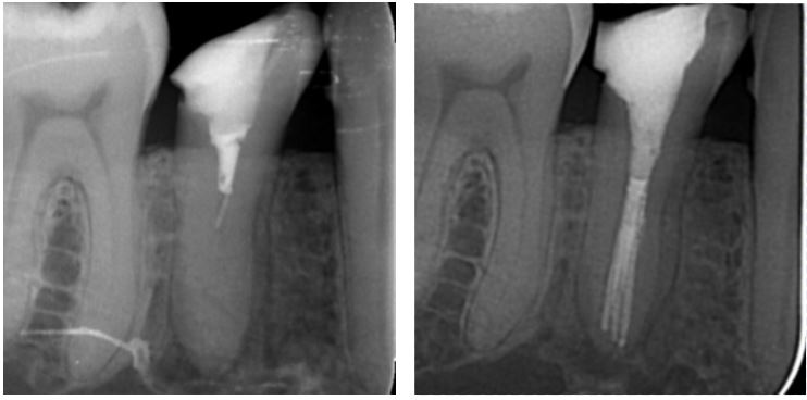
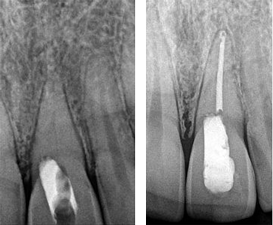
CASE 2
A young patient with internal resorption of the palatal canal. Thermoplastic obturation allowed for a complete fill of the defect prior to restoring the access cavity with a deep base of GIC and a bonded restoration.
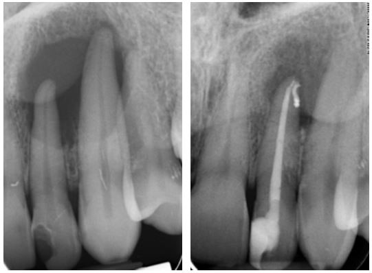
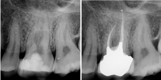
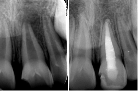
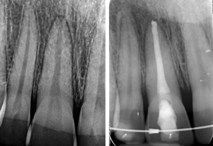
Calcified Pulp Canals
CASE 1
RCT was attempted by the referring dentist on the LL1 and LL2. As the pulp canals were obliterated, the canals were not located and the patient was referred. Henk located the canals completed RCT by placing GIC plugs over the gutta percha and temporizing the access.
CASE 2
The challenges of performing endodontic treatment on this UL1 was recognised by the referring dentist and reccommended Henk perform the treatment. The post- treatment radiograph shows conservative access filled with GIC and with minimal loss of tooth material.
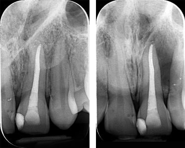
Healing of Large Periapical Lesions with Non-Surgical Treatment
CASE 1
Whilst large periapical radiolucencies are more likely to be radicular cysts than granulomas, they generally respond well to non-surgical treatment.
This carious UL2 developed a draining fistula bucally and swellings both bucally and palatally, thus representing a “through and through” defect i.e. loss of both the buccal and palatal cortices. Conventional RCT was performed and the 12 month review radiograph shows excellent osseous healing underway.
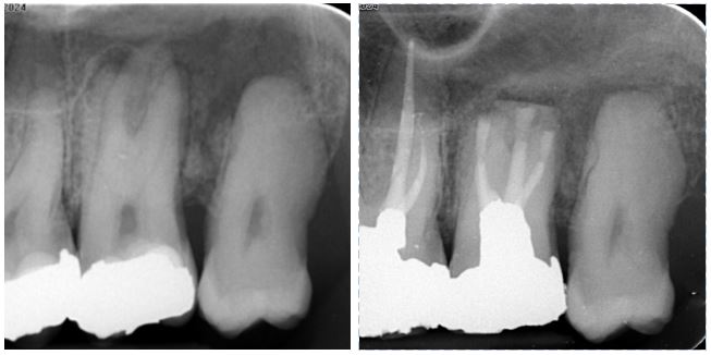
CASE 2
This case was referred due to buccal swelling associated with the lower incissors. The LL2 was suspected to be the the source of the problem as it had grade II mobility. The periapical radiograph shows a large radiolucency with significant loss of bone between LL2 and LL3. The coronal CBCTslice (bottom right) shows the lesion to extend from LR1 to LL3. Henk determined LL2 to the endodontic source of the lesion. CBCT helped to exclude root fractures and the sagittal slice of LL2 (bottom left) shows this tooth to have buccal and lingual canals. Conventional RCT was performed and the six month review radiograph (top right) shows almost full resolution of the lesion
CASE 3
The root treated and crowned UR1 and UR2 were the source of a large radiolucency over their apices, with associated buccal and palatal swellings. A history of trauma indicated a CBCT scan to help exclude root fractures. This shows significant loss of the buccal cortex (reconstructed CBCT, bottom left), as well as cortical erosion palatally.
Inter-appointment calcium hydroxide dressings were placed for 12 weeks. The signs and symptoms resolved and the case was completed. The post-treatment radiograph (top right) shows early, but clear reduction in the size of the lesion and new osteoid bone deposits centrally after just 12 weeks.
Intentional Replantations
The outcomes for intentional replantations is generally very good indeed. This treatment modality is used where conventional apical surgery is contraindicated or high risk. This UL7 was the source of a palatal draining fistula and large radiolucency over the UL7 and UL8. Signs and symptoms did not resolve after shaming, cleaning and calcium hydroxide dressing. The decision to remove it, perform apical resection and place retrograde MTA plugs was taken and the tooth replanted without any splinting. Signs and symptoms resolved shortly therafter and the 4 month review radiograph shows excellent periapical resolution underway.
CASE 2
This root treated and post -crowned LR5 developed a symptomatic apical periodontitis. The patient was an elderly lady who couldn’t lollerate a typically lond re-RCT appointment. The decision to perform a 15 minute replantation procedure, as in case 1 above, was taken. Again no splint was used after replanting. The 12 month review radiograph shown a new PDL becoming established apically.
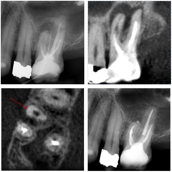
Perforation Repairs
CASE 1
Iatrogenic pulp floor perforations are not uncommon. Fortunatley, they generally respond very well to treatment. This upper 6 shows furcal bone loss due to a frank perforation a few months earlier. Henk performed RCT, perforation repair with MTA and an amalgam Nayyar core. The six month review radiograph shows furcal bone regeneration.
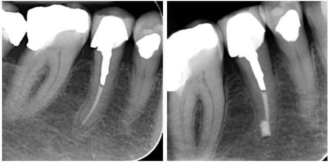
CASE 2
Root perforations are quite uncommon but can occur with endodontic instrumentation or post-space preperation. This lower 6 has significant furcal bone loss due to perforation of the distal root. The canals were obturated with bioceramic materials, and the perforation was repaired with bioceramic putty before temporizing.
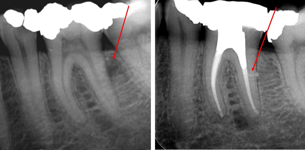
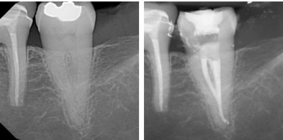
Lateral Canals
CASE 1
The lateral radicular radiolucency on the distal root of this lower 6 was the cause of of a periradicular abscess. The three month review radiograph shows complete healing of the lesion with the associated lateral canal filled. Bioceramics were used in this case. While bioceramic sealers are superb, they are not very radiopaque.
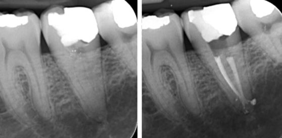
CASE 2
This upper 5 shows good periapical healing at six month review. The magnified periapical view shows lateral canals filled with bioceramic sealer.
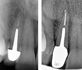
Open Apices
CASE 1
Immature teeth with endodontic infections are, generally, treated using an “apexification” procedure to provide an apical barrier. For this upper central in an 8 year old, an MTA apical plug was placed and the rest of the canal backfilled with Thermoplastic g.p. and the access restored with GIC. The 6 month review radiograph shows periapical healing underway
CASE 2
This upper central incissor has a fairly mature root, however the apex was gauged to a size too large for predictable sealing with g.p. and sealer. Bioceramic putty was used for an apical plug and the remaining canal filled with bioceramic g.p.and sealer. A base of GIC was placed at the ECJ level im preperation for future whitening and the access temporized with IRM, as seen on the 6 month review radiograph.
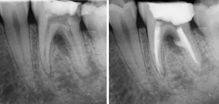
“C” Shaped Canals
CASE 1
C shaped canals are mostly found in lower second molars, however, they are also found in upper molars and rarely in lower premolars. There is one root with merging canals making them difficult to shape, clean and fill well. This lower 7 was completed with a base of GIC over the g.p. prior to placing a core and crown.
CASE 2
Similar to case 1 above, except after the canals merge, they seperate and have multiple portals of exit.
Apical Micro-Surgery
Cases with external root resorption
Here a large lesion is associated with missed anatomy and a leaking silver point. The crown was removed and the tooth re-treated.
A Nayyar core and temporary crown were placed. Healing was demonstrated at 9 months so that the referring dentist could confidently place a new crown.
The pre-treatment radiograph shows very large lesions on this Lower 6, it also had with a draining sinus.
Management included long term dressing with an iodoform paste. Healing of these lesions is demonstrated after only three months when the next radiograph was taken at completion of the treatment.
A Lateral canal has been filled in coronal half of the mesial root system.
Cases with internal root resorption
The initial radiograph was taken on completion of the treatment and shows a great deal of bone loss on the lateral aspect of the UL1.
Almost full resolution is seen between UL1 and UL2 at the 12 month follow-up.
Cases with perforation defects
Post removal, re-treatment and healing at 12 month follow-up.
Replantation Cases
A referring dentist separated an instrument deep in this upper premolar.
After removing it and completing RCT a Nayyar core was placed so the referring dentist could easily prepare a crown.
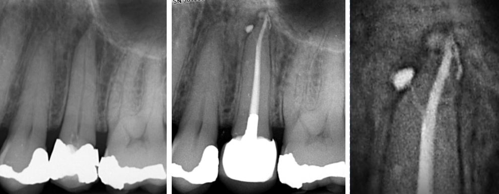
Apical Micro-Surgery
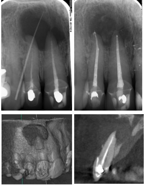
Obliterated canals are notoriously difficult to locate, even with a microscope.
Here a referring dentist could not locate the canal in this UL1 and rightly referred the case to Henk before perforating.
Henk located the canal and completed treatment using ISO files so as not to weaken the tooth further.
ENDODONTIC MICROSURGERY
Call Us for an Appointment
Make An Online Enquiry